
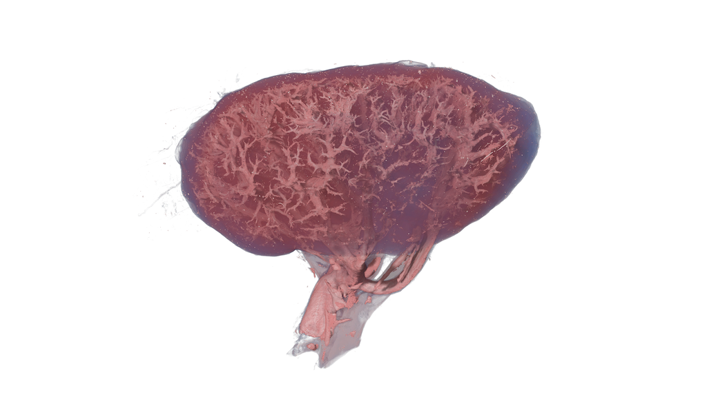
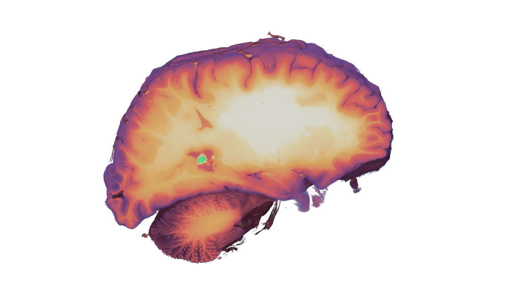
Turning ultra‑high‑resolution biology into explorable understanding
This exhibit turns real scientific data into an explorable experience. Built on authentic imaging and developed with leading research partners, it combines scientific accuracy, thoughtful design, and intuitive interaction—making complex subjects accessible without reducing their depth.

Exhibit at a Glance
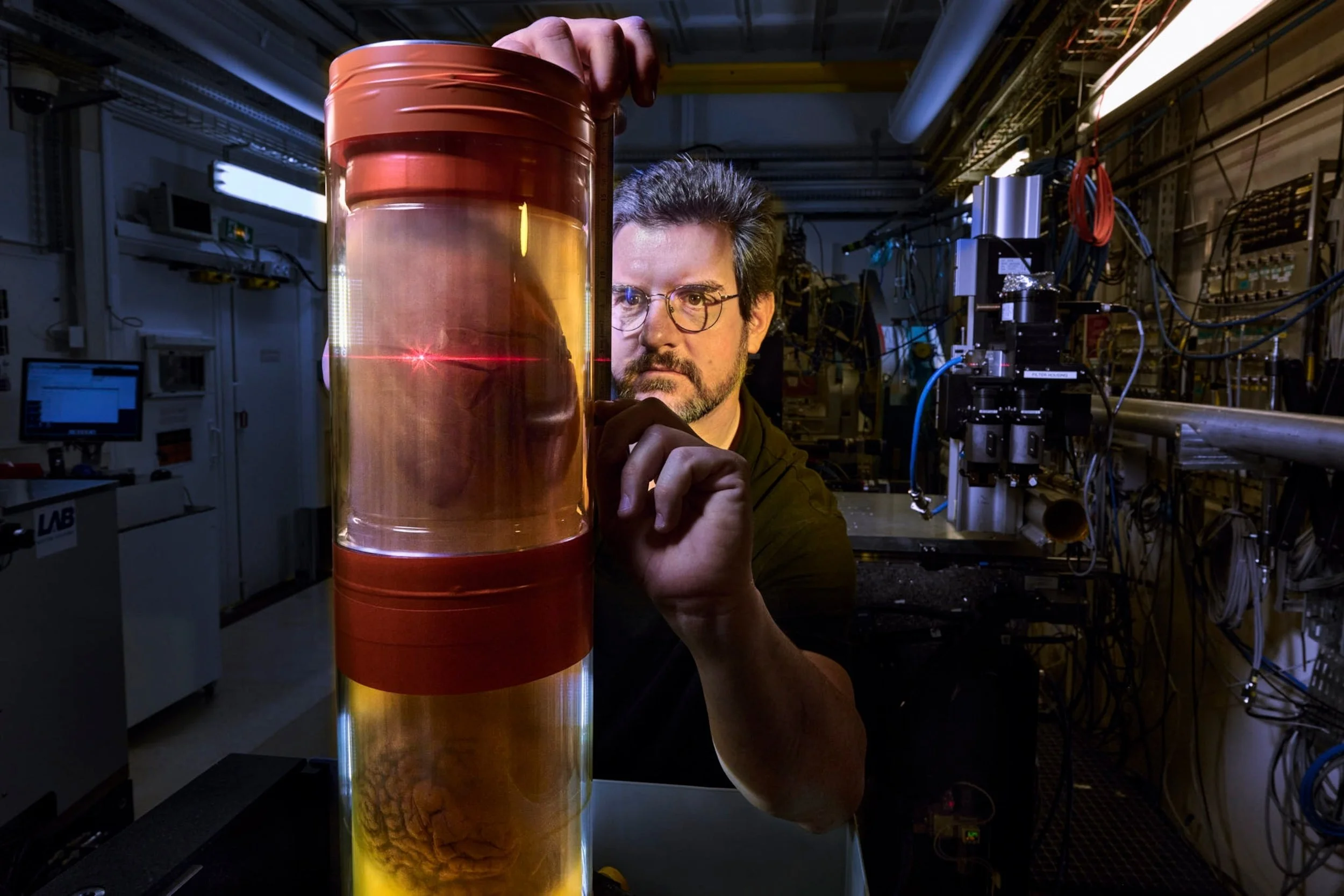
World‑leading imaging data – Built on ultra‑high‑resolution synchrotron scans from the European Synchrotron Radiation Facility (ESRF)
HIP‑CT technology – Uses Hierarchical Phase‑Contrast Tomography to reveal anatomy from whole organs down to near‑cellular detail
Explorable human organs – Interactive 3D exploration of brain, lungs, kidneys, spleen, and heart tissue
From organ to microstructure – Enables seamless zoom from full anatomy to microscopic features invisible to conventional medical CT
Authentic research data – Visitors explore the same datasets used by scientists and medical researchers
Guided yet open‑ended – Rich annotations support learning while allowing free, curiosity‑driven exploration
Health and disease insight – Includes real pathological cases such as COVID‑19 lung damage
Designed for education and outreach – Suitable for museums, science centres, universities, and public learning environments
Featured in the exhibit
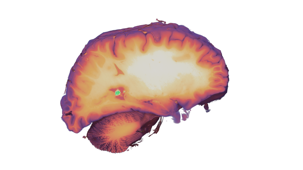
Human Brain (HiP‑CT synchrotron scan)
Ultra‑high‑resolution scan showing detailed brain structures, including grey and white matter, lobes, and internal regions.
Data by: ESRF European Synchrotron Radiation Facility (ESRF) / Human Organ Atlas
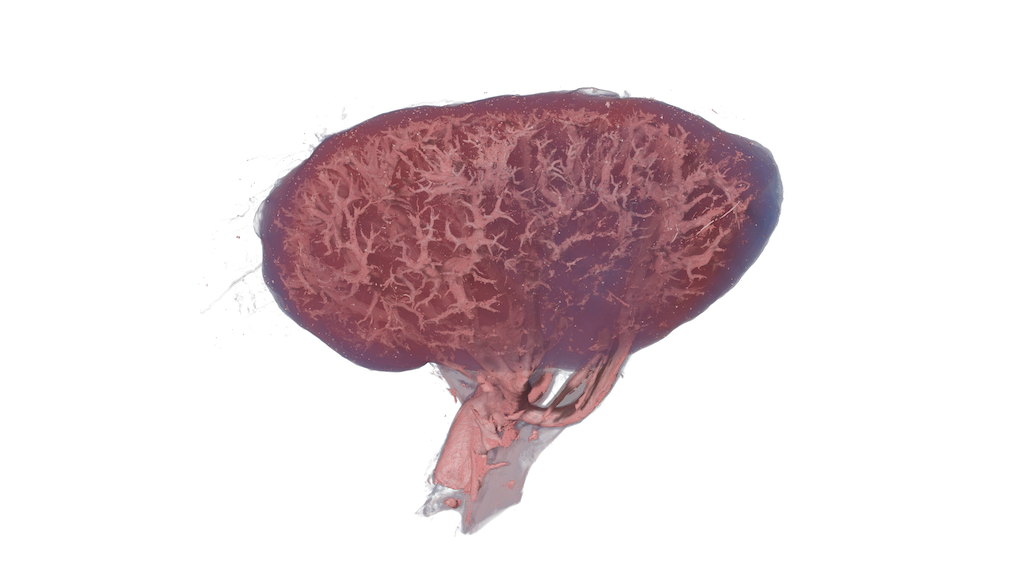
Left kidney
High‑resolution scan revealing internal structures such as nephrons, renal pyramids, and filtration systems.
Data by: ESRF European Synchrotron Radiation Facility (ESRF) / Human Organ Atlas
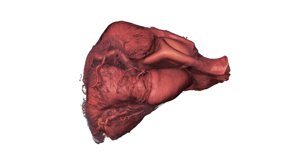
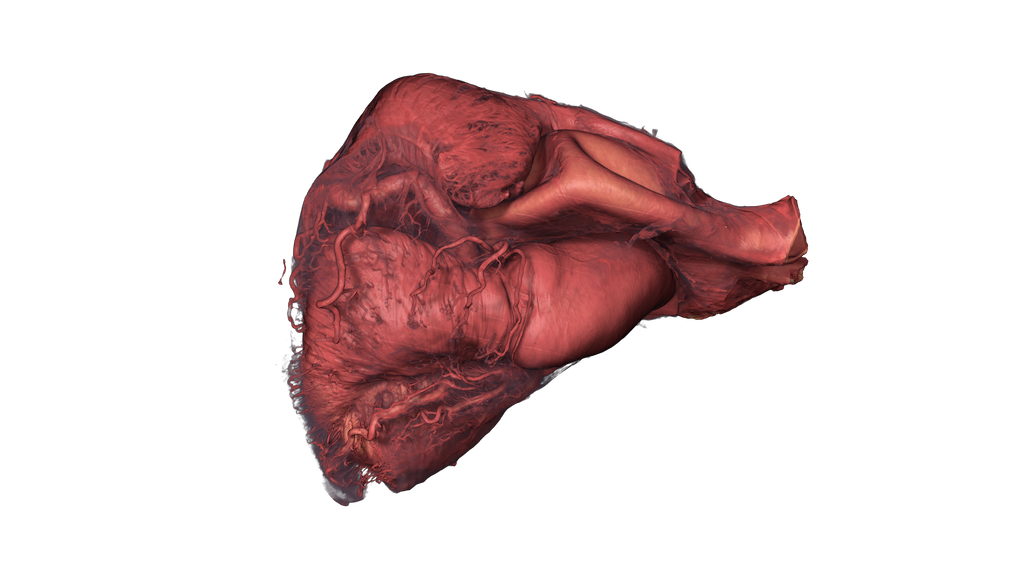
Heart (multi‑scale samples)
High‑resolution datasets showing internal cardiac structures at different levels of detail.
Data by: ESRF European Synchrotron Radiation Facility (ESRF) / Human Organ Atlas
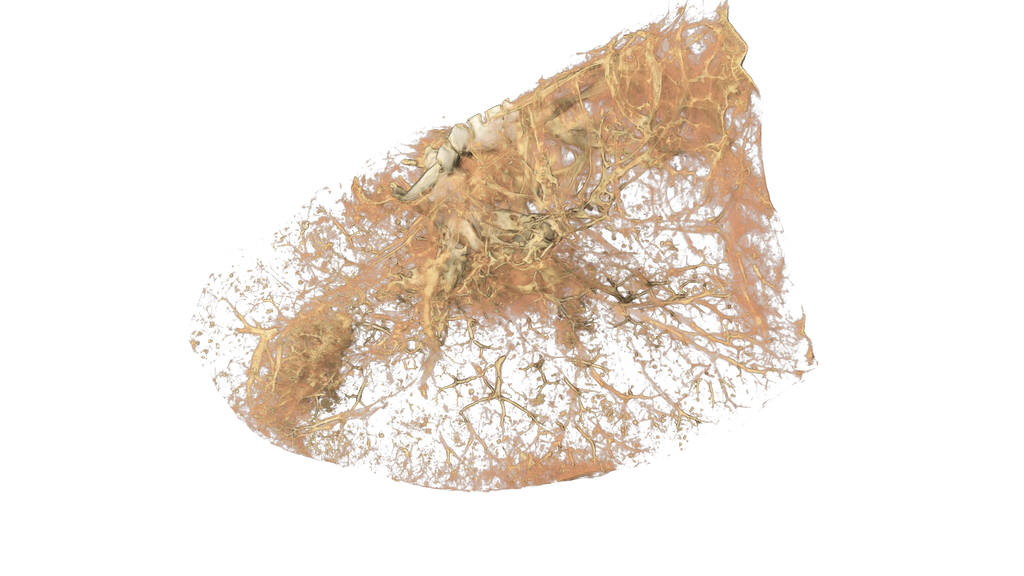
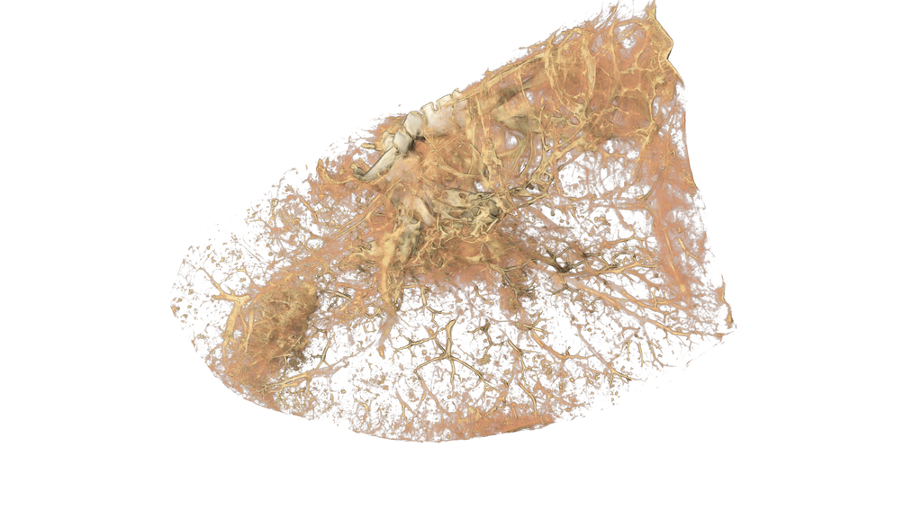
COVID‑19 Lung (Upper Left Lobe)
Synchrotron CT scan of lung tissue showing structural damage caused by COVID‑19 (ARDS), including alveolar and vascular changes.
Data by: ESRF European Synchrotron Radiation Facility (ESRF) / Human Organ Atlas
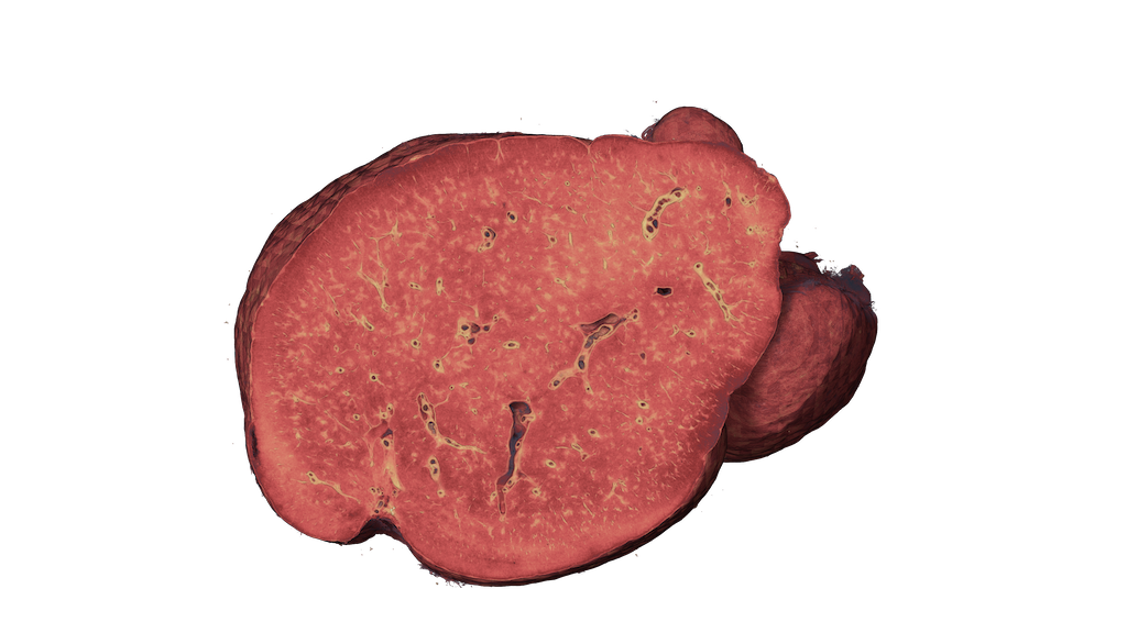
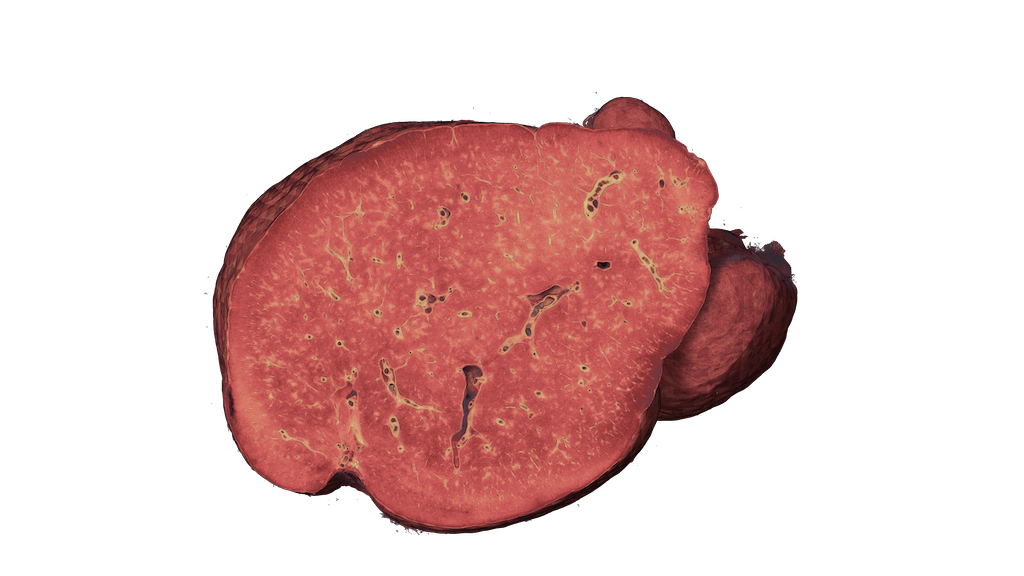
Spleen
Detailed scan of lymphatic and blood‑filtering structures, including red and white pulp.
Data by: ESRF European Synchrotron Radiation Facility (ESRF) / Human Organ Atlas