
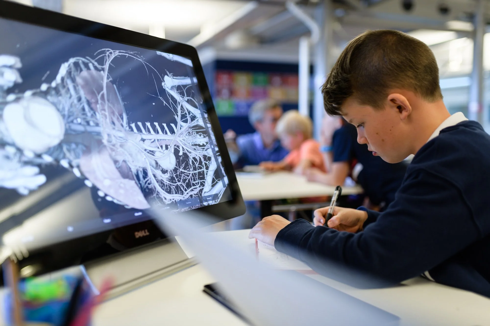
Explore how real CT data, 3D modelling, and patient‑specific implants come together—revealing the future of personalised medicine through interactive, explorable anatomy.

Exhibit at a Glance
Real clinical imaging data – Built on authentic CT scans of living patients, developed in collaboration with medical experts
From scan to solution – Shows the full workflow from CT imaging and 3D modelling to patient‑specific implants and treatment
Patient‑specific medicine – Demonstrates how custom‑made 3D‑printed implants are designed to match individual anatomy
Real medical cases – Covers conditions such as Pectus Excavatum, Poland Syndrome, airway stenosis, and maxillofacial reconstruction
Explorable surgical insight – Enables users to explore anatomy, pathologies, and implant placement through interactive 3D visualisation
Low‑invasive treatment focus – Highlights modern surgical approaches that reduce patient trauma and improve outcomes
High scientific credibility – Grounded in real medical data, without illustrations or simplification
Featured in the exhibit
POLAND SYNDROME
CT scan acquisition of a living patient suffering of a Poland Syndrome congenital disease. Poland Syndrome is a partial or total lack of the Pectoralis Major muscle. It can be corrected by inserting a 3D custom-made implant trough surgery.
Data by IMA Solutions SARL, France
PECTUS EXCAVATUM
CT scan acquisition of a living patient suffering of a Pectus Excavatum congenital deformation. Pectus Excavatum is a deformation of the ribs and sternum. With a frequency of 1 over 300 births, it is the most common thoracic congenital deformation. This patient underwent a surgery to place a 3D custom-made implant to correct the deformation.
Data by IMA Solutions SARL, France
AIRWAY STENOSIS DISEASE
Chest CT scan with contrast of a living patient suffering from an airway stenosis. The patient cannot breathe normally because of a reduction of the section of the airway. Treatment consists in implanting an airway stent to get a normal airway section.
Data by IMA Solutions SARL, France
MANDIBULAE RESECTION - CANCER
Face CT scan of a living patient. The mandibulae bone as been partially resected to remove a cancer tumor. Patient will undergo a surgery to reconstruct the missing mandibulae part.
Data by IMA Solutions SARL, France
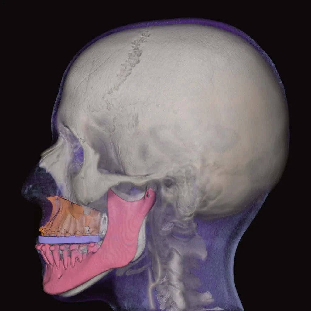
MAXILLOFACIAL - PROTRUDING MANDIBULAE
Face CT scan of a living patient. The mandibulae bone is protruding meaning a bad alignment of upper and lower teeth. The patient will undergo a surgery to realign correctly the mandibulae with the maxillary bone.
Data by IMA Solutions SARL, France