
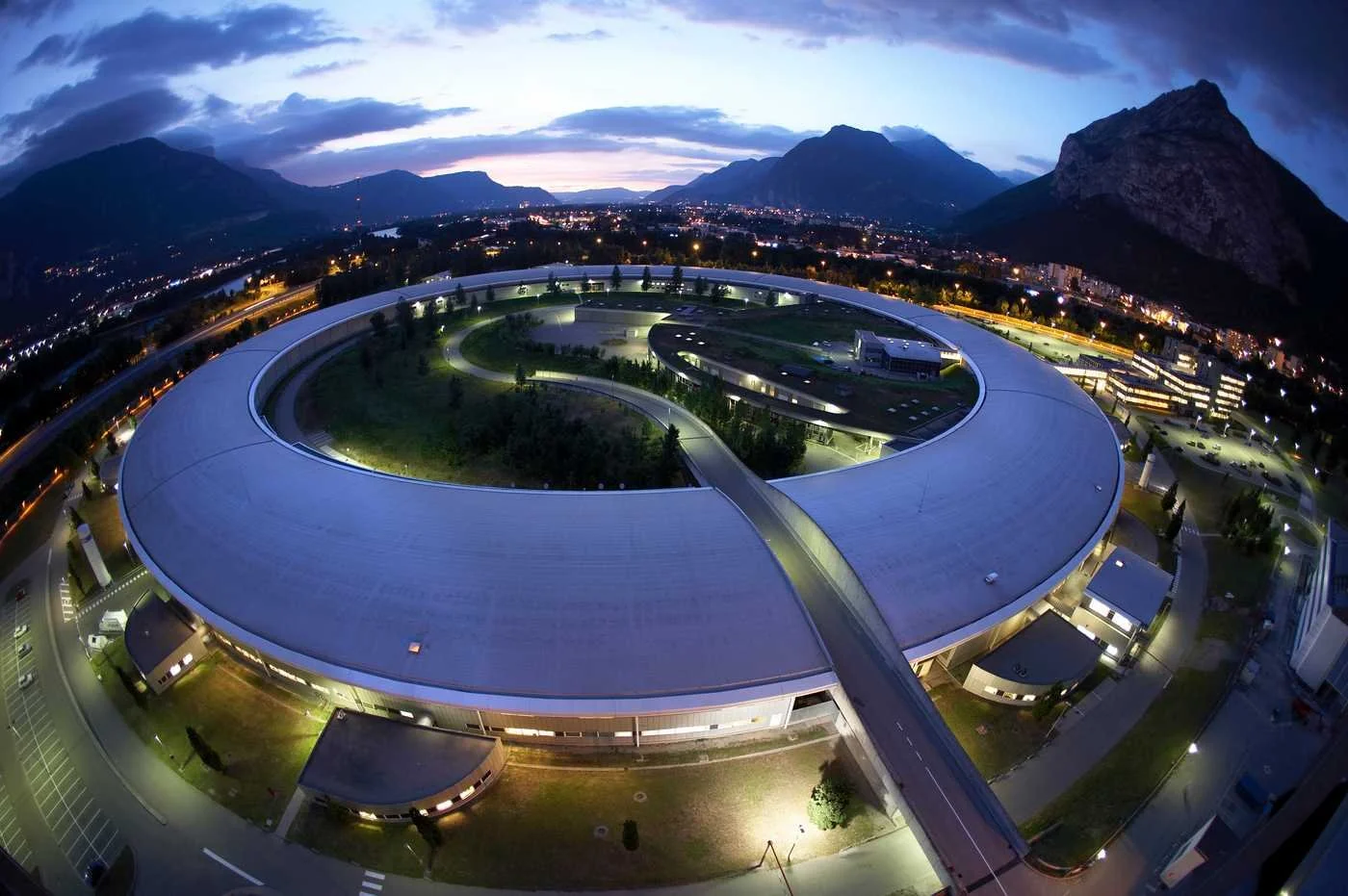
European Synchrotron Radiation Facility
Making the world’s highest‑resolution anatomy scans accessible
Storyvoxel partnered with the European Synchrotron Radiation Facility (ESRF) to make ultra‑high‑resolution synchrotron CT scans of human anatomy explorable through interactive streaming services and on‑site installations.
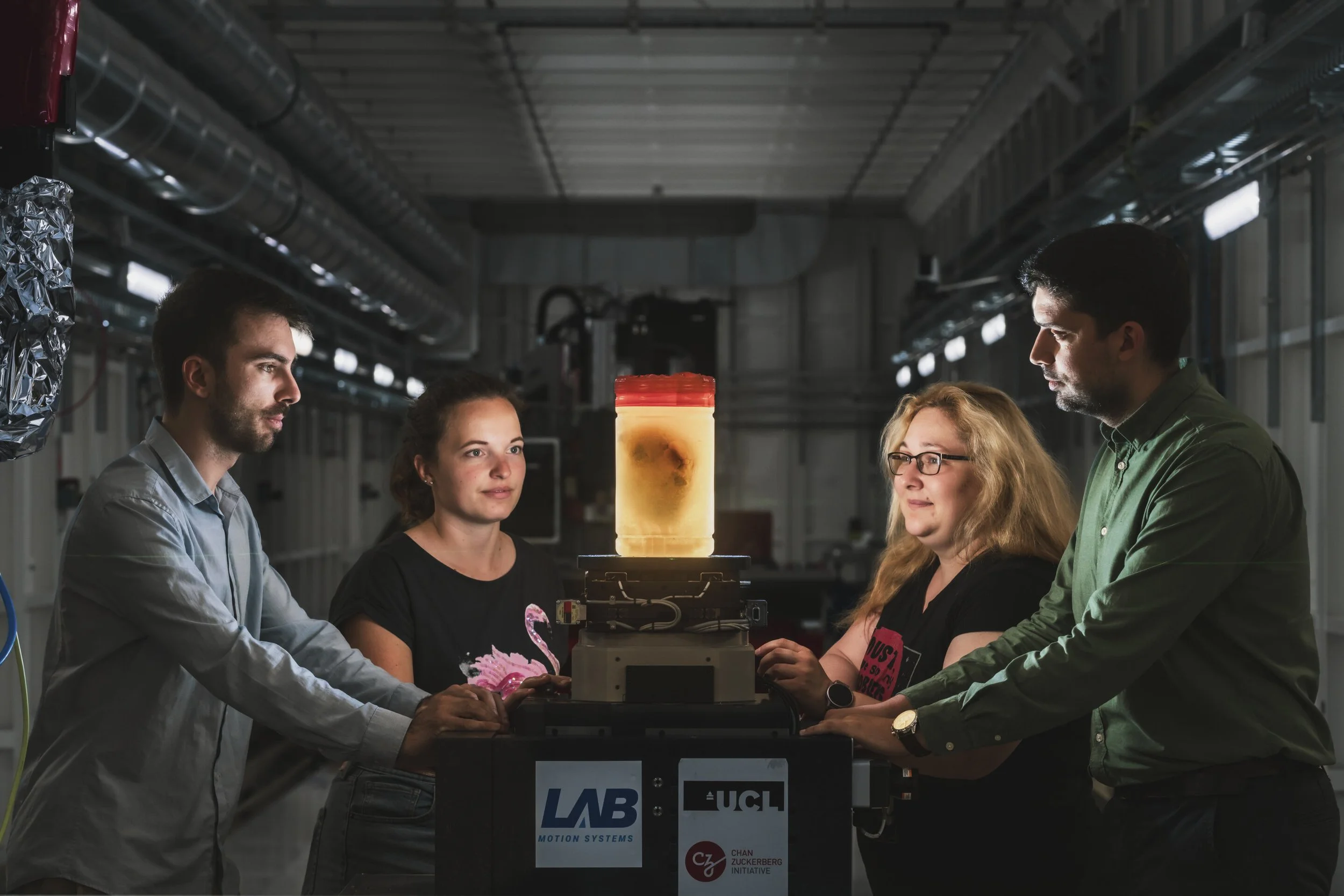
The project is part of the UCL‑ESRF Human Organ Atlas and is based on some of the most detailed 3D scans of human organs ever produced. Using Storyvoxel’s interactive technology, complex volumetric datasets are made accessible for education, research communication, and public engagement—without reducing complexity.
The challenge
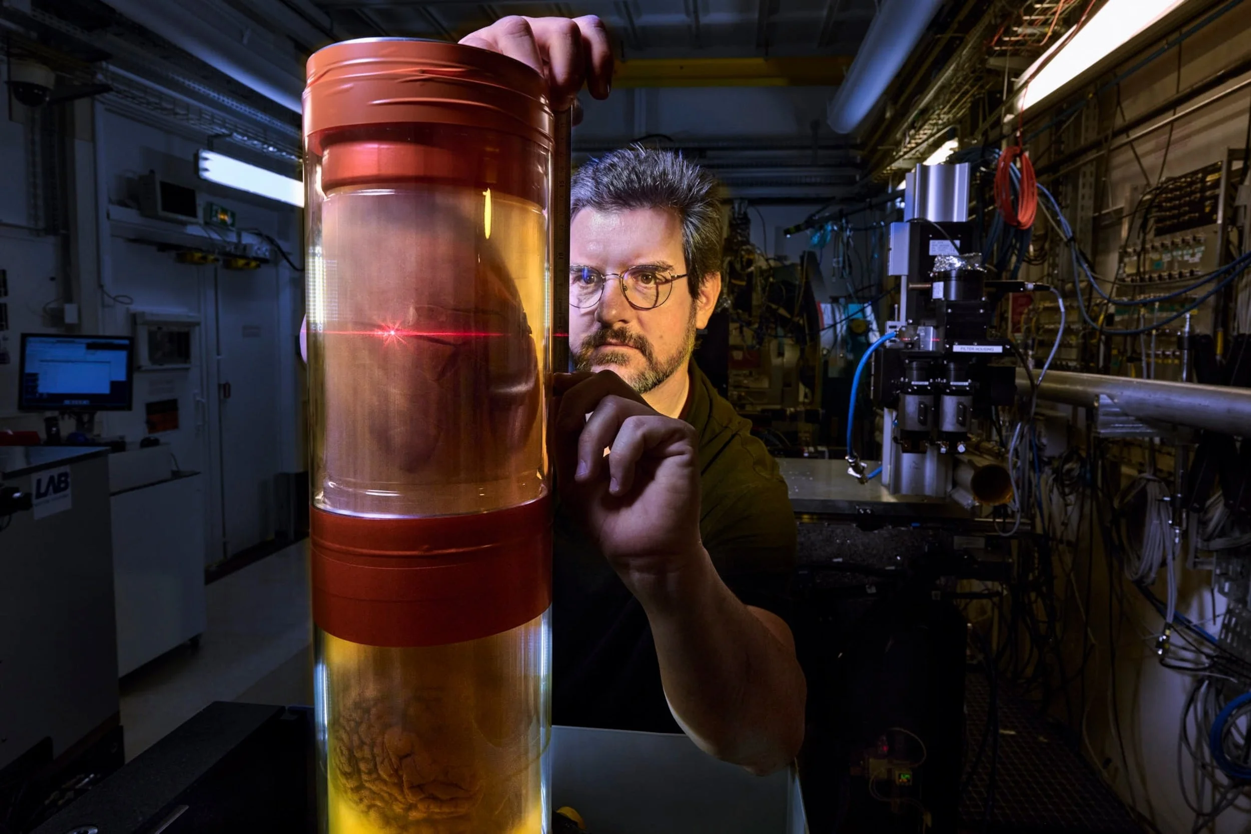
Synchrotron CT imaging produces extraordinarily detailed 3D datasets, capturing structures far beyond the resolution of conventional medical CT. While scientifically invaluable, these datasets are extremely large and complex, making them difficult to share, explore, and communicate beyond specialist environments.
ESRF needed a way to provide access to real synchrotron data that would work both online and in public‑facing spaces—allowing users to explore the data directly without requiring specialist software, high‑end hardware, or large data downloads.
The Solution
Storyvoxel delivered a streaming solution that lets anyone explore the Human Organ Atlas scans directly in a web browser. The full datasets stay on the server and are rendered there, and only the interactive view is streamed to the user. This means there is nothing to download, no specialist software to install and no need for powerful hardware. A laptop, tablet or phone is enough to zoom from a whole organ down to fine anatomical detail.
The same data is also available on site. In the ESRF visitor centre, an interactive installation lets visitors explore the organ scans hands-on, using the same intuitive interaction as the online service.
The data
The dataset is captured using HiP‑CT (Hierarchical Phase‑Contrast Tomography), a novel synchrotron imaging technique developed through a pan‑European collaboration between University College London (UCL) and ESRF.
HiP‑CT enables full human organs to be scanned at resolutions up to 100 times higher than conventional medical CT, revealing anatomical detail across multiple scales.
The result
Advanced CT imaging generates highly detailed and information‑rich datasets, but these are often difficult to communicate beyond specialist users. Static images, reports, or expert‑only tools limit access, making it harder to engage collaborators, students, partners, and visitors in meaningful dialogue around the data.
DTU’s 3D Imaging Centre wanted a way to open up access to real CT data directly in the lab environment—supporting demonstrations, collaboration, and communication with diverse audiences, while preserving scientific depth and fidelity.
“With the streaming service from Interspectral we now have the possibility to visualize our complex data and stream high-resolution 3D scans of human organs directly on the Human Organ Atlas website”
— Andy Götz, ESRF
Key outcomes
Ultra‑high‑resolution CT data made explorable beyond specialist tools
Shared, interactive access to real synchrotron imaging data online and in the visitor centre
Improved communication and understanding across research, education, and public engagement
Preserved scientific depth through layered, interactive exploration