
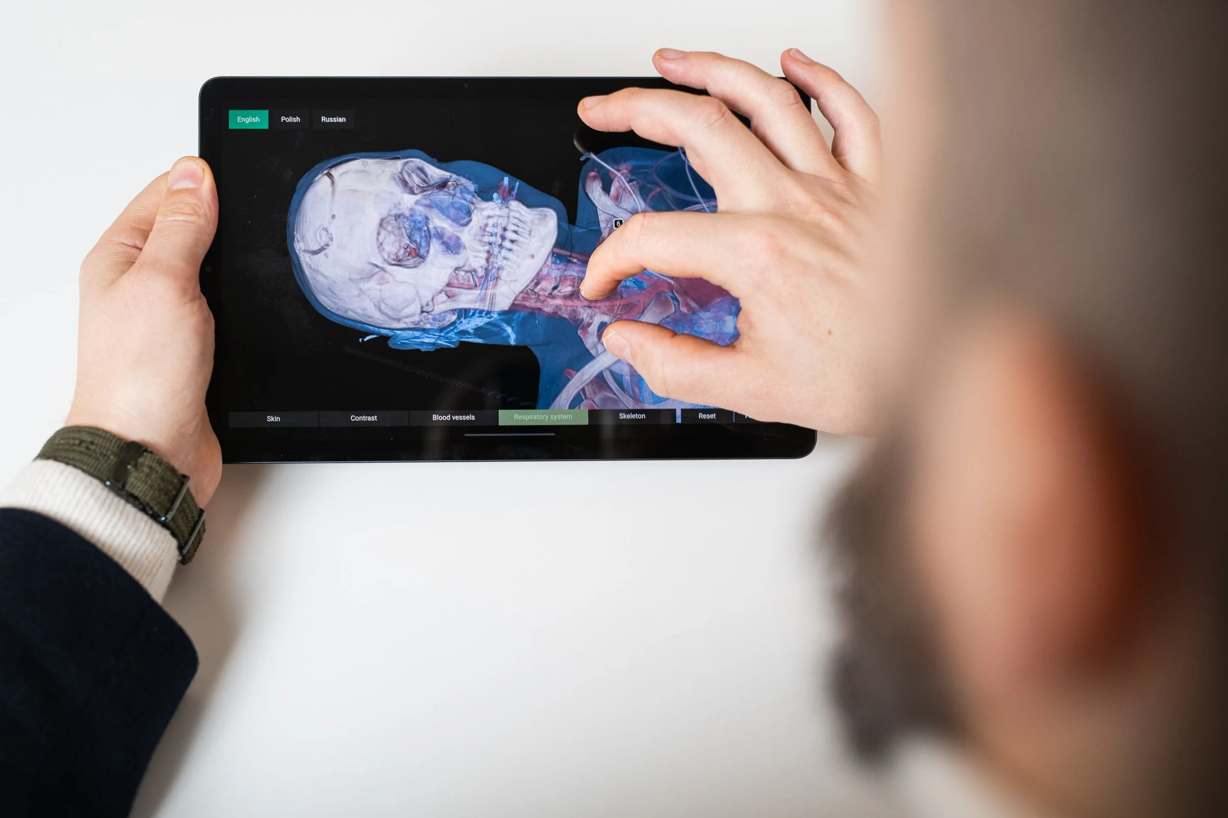
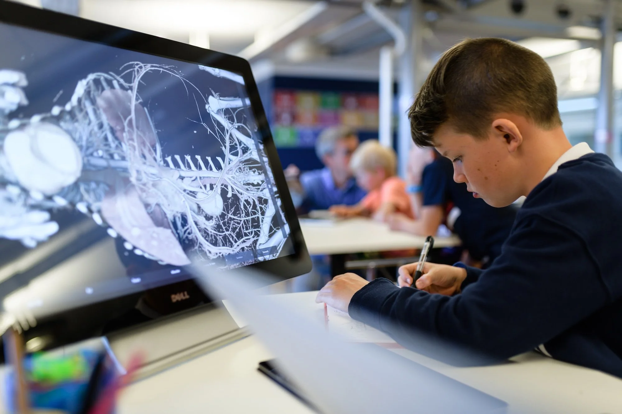
Explore the human body in unprecedented detail through an interactive, data‑driven anatomy experience built on real scientific imaging.
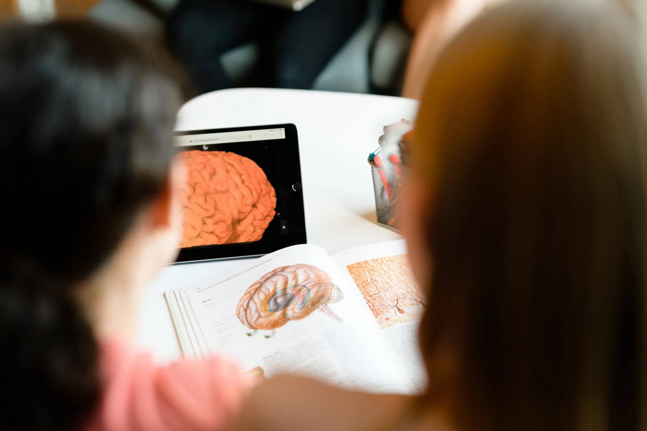
Exhibit at a Glance
Real medical imaging data – Built on authentic CT datasets from clinical and research sources
Explorable human anatomy – Interactive, layered exploration of the human body through real cases
From health to pathology – Covers trauma, stroke, aging, medical implants, and advanced brain anatomy
Learning through investigation – Enables users to explore causes, effects, and anatomy via direct interaction
Virtual autopsy experience – Supports deep, non‑destructive exploration similar to professional medical tools
High scientific fidelity – Preserves clinical accuracy without relying on illustrations or synthetic models
Flexible use – Suitable for public exhibitions, education, and classroom learning environments
Featured in the exhibit
FULL BODY SCAN - TRAFFIC ACCIDENT
A scan of a woman who died in a road accident. The examination provides a quick overview of the dead woman's anatomy and can reveal the cause of death-in this case, a broken neck. There are also a number of visible fractures to the jaw, on the right foot, left tibia and fibula, the right knee, several pelvic fractures, and the left neck of the femur.
Data by Linköping university, Sweden
FULL BODY CT SCAN - SIGNS OF AGING
Scan of a man in his fifties in physically good health. An example of natural aging of the body is seen in the spine where the height between the first and second vertebra is reduced. You can also see calcification in the vertebrae, also a result of aging.
Data by Linköping university, Sweden
BIG BRAIN ATLAS
Brain Atlas is used by other scientists investigating specific parts of the brain. In this dataset, you will be able to explore the highest resolution 3D Brain Atlas yet made of a human brain and learn more about the brain.
STROKE PATIENT
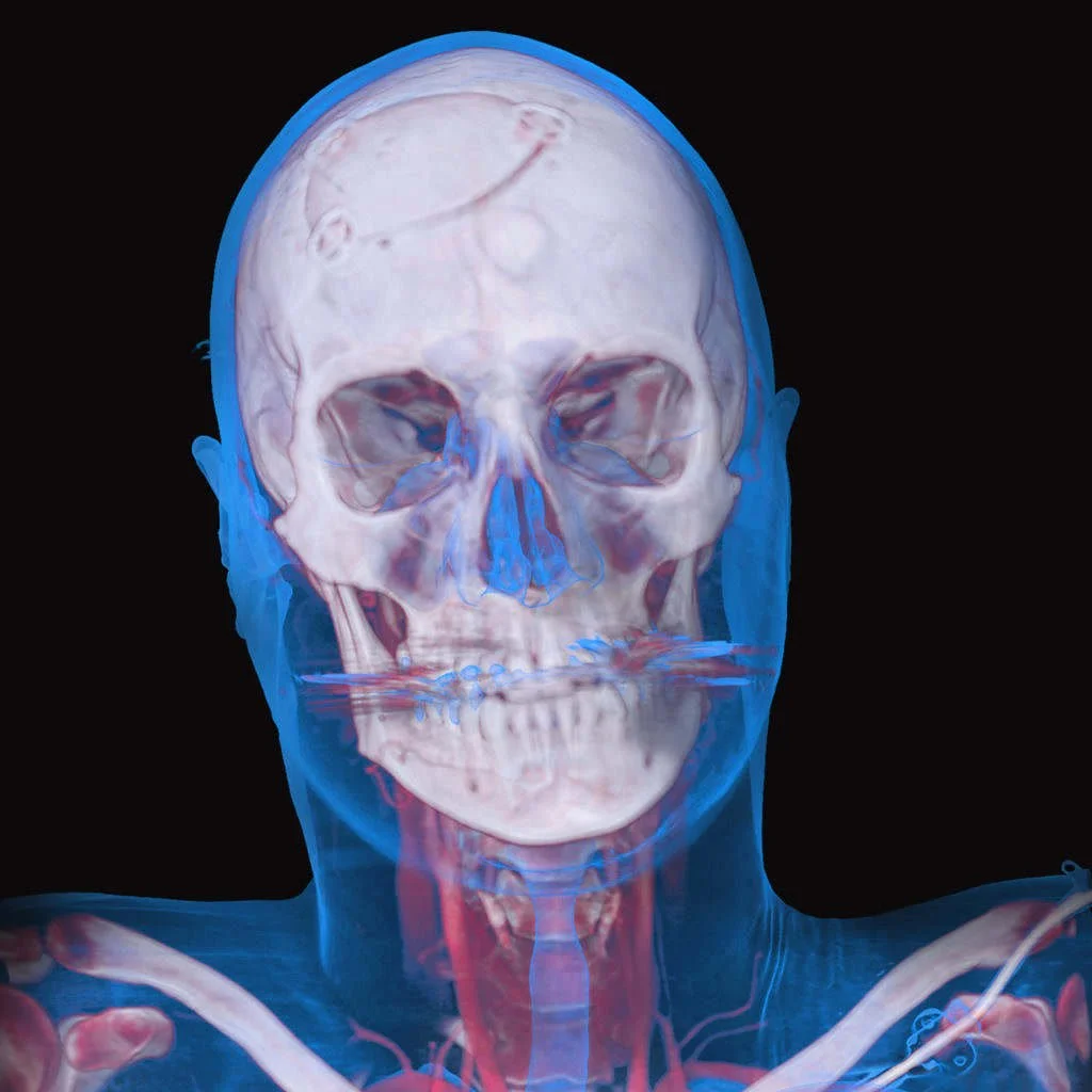
A CT scan of a living patient who has been treated for stroke. The patient has undergone surgery for a ruptured aneurysm in a small blood vessel in the brain using a procedure called "clipping", which simply put means the burst blood vessel is repaired using a small metal clip.
Data by Linköping university, Sweden
ARTIFICIAL HEART PUMP
The heart pump fits neatly into a cavity below the heart next to the diaphragm. It connects to the left ventricle, where the blood leaves the heart. This particular type of pump is used where heart function has all but failed and conventional medical treatment is unable to help.
Data by Linköping university, Sweden